Quick answer
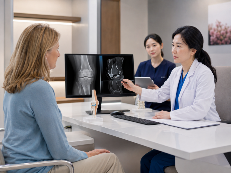
An X-ray shows the bones and the space between them, which is why it is used to grade knee osteoarthritis. An MRI shows the soft tissues an X-ray cannot see: the cartilage itself, the meniscus, the ligaments, and swelling inside the bone. MRI is most useful when its answer could change the treatment plan, and its findings need to be read alongside your symptoms, because many knees show changes on MRI without causing pain.
Key takeaways
- A standing X-ray is the usual first test and grades osteoarthritis from the joint space and bone changes.
- MRI adds detail on cartilage defects, the meniscus and bone marrow lesions, which matters when a cartilage-preserving treatment is being considered.
- MRI findings are common in knees that do not hurt, so a report is interpreted together with an examination, not on its own.
What an X-ray shows
A knee X-ray is a picture of bone. Cartilage is invisible on it, but its thickness can be inferred from the gap between the thigh bone and the shin bone. When cartilage wears, that gap narrows. The X-ray also shows bony spurs at the edges of the joint and hardening of the bone just beneath the cartilage.
These three features form the basis of the Kellgren–Lawrence scale, which grades osteoarthritis from 0 to 4.1 The X-ray should be taken while you stand, because the joint space looks wider when the leg is not carrying weight. A full-length standing X-ray of both legs can also show whether the knee is bow-legged or knock-kneed, which affects how load passes through the joint.
X-rays are quick, inexpensive and good at what they do. Their limit is that they show the result of cartilage loss, not the cartilage itself. Two knees with the same joint space can have very different cartilage surfaces, and early damage may not show at all.
There is also a well-known mismatch between X-ray findings and symptoms. A review of the published studies found that among people with knee pain, the proportion who had osteoarthritis on X-ray ranged from 15% to 76%, and among people with X-ray osteoarthritis, the proportion with pain ranged from 15% to 81%.2 The X-ray is one piece of the picture.
What an MRI adds
MRI uses a magnetic field rather than radiation and shows soft tissue in detail. For the knee, it can show:
- Cartilage: the size, depth and location of a defect, including whether it reaches the bone beneath.
- Meniscus: tears, and whether the meniscus has been pushed out from between the bones.
- Ligaments: such as the anterior cruciate ligament, which affects stability.
- Bone marrow lesions: areas of fluid and stress inside the bone just below the cartilage.
- Joint fluid and lining: swelling and inflammation of the joint lining.
Bone marrow lesions deserve a special mention because an X-ray cannot see them at all. In a study of 401 people with knee osteoarthritis, these lesions were found in 77.5% of those with knee pain and in 30% of those without pain.3 They help explain why two knees with the same X-ray grade can feel so different.
For treatments that aim to preserve or repair cartilage, MRI is often essential. The size and depth of a defect decide whether a focal cartilage procedure is possible at all, and the state of the meniscus and alignment often decide whether it is likely to last.
Why MRI findings need careful reading
MRI is sensitive. That is its strength, and also the reason its reports can alarm people unnecessarily.
In a community study of 710 adults aged 50 and over whose X-rays showed no osteoarthritis at all, 89% had at least one abnormality on MRI.4 Cartilage damage appeared in 69%, bony spurs in 74%, and bone marrow lesions in 52%, and these findings were common whether or not the person had knee pain. Meniscus tears are similar: in the same age group, most people found to have a tear on MRI had no knee pain, aching or stiffness in the previous month.5
Think of an MRI report like a detailed home inspection. A careful inspector lists every crack, but only some of them matter for whether the house is safe to live in. The job of the orthopedic review is to decide which findings explain your symptoms and which are part of normal aging.
This is also why an MRI is usually requested to answer a specific question, such as whether a cartilage defect is suitable for repair, rather than as a routine first test for knee pain.
When each test is usually used
For most people with suspected knee osteoarthritis, a standing X-ray and a physical examination are the starting point. An MRI is more likely to be recommended in these situations:
- The knee locks, catches or gives way, suggesting a meniscus or ligament problem.
- Pain seems out of proportion to a mild X-ray.
- A cartilage-preserving treatment is being considered and the defect needs to be measured.
- Symptoms started suddenly after an injury.
- The diagnosis is uncertain and other causes need to be ruled out.
If you already have an MRI from home, it is often worth having it reviewed before repeating it. A recent scan of good quality can usually be used for planning, although a new scan may be needed if the old one is outdated or the images are unclear.
How imaging is handled at St. Luke’s Hospital
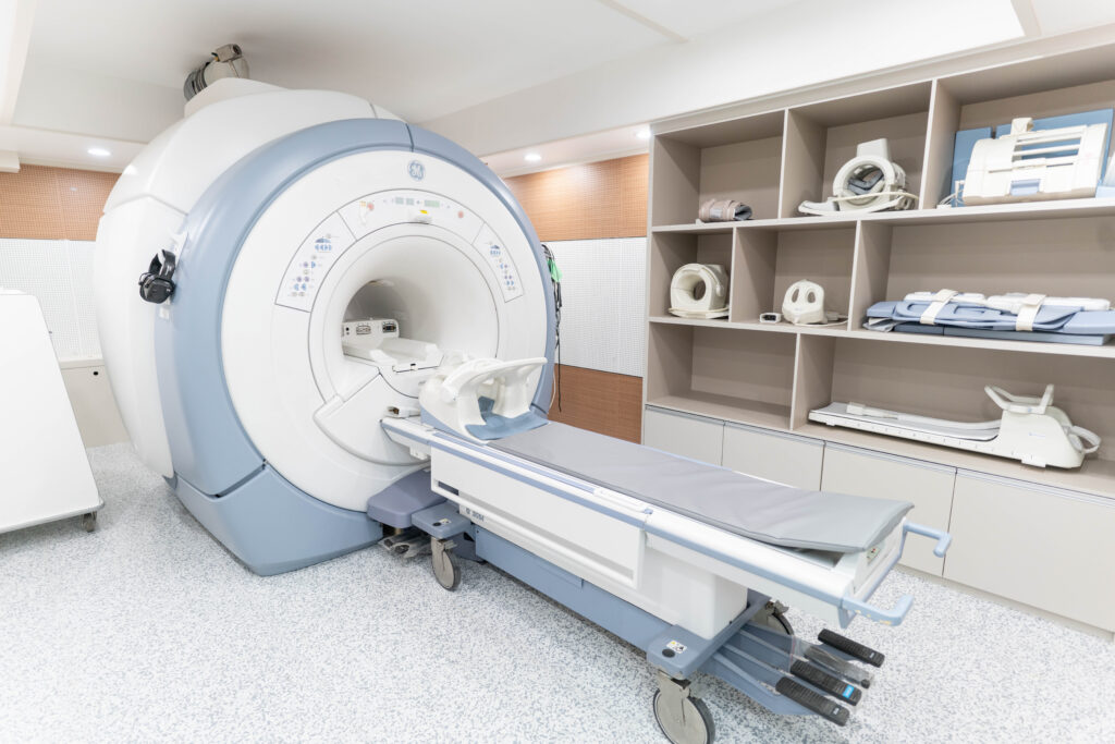
X-ray and MRI are performed on site at St. Luke’s Hospital, and images are interpreted by a radiologist based at the hospital. That means imaging, reading and the orthopedic consultation can often take place close together, which is useful when your time in Seoul is limited.
Our MRI uses SwiftMR, an AI image-reconstruction software by AIRS Medical that is cleared by the U.S. Food and Drug Administration. It is designed to produce clearer images in a shorter scan. The software improves the image; the diagnosis still comes from the radiologist and the orthopedic specialist who read it together with your examination.
If you are overseas, you can send your existing X-rays and MRI for a preliminary review before deciding whether to travel. For an overview of how imaging fits into treatment choices, see what treatment fits each stage of knee osteoarthritis.
Frequently asked questions
Do I need an MRI to be diagnosed with knee osteoarthritis?
Usually not. Knee osteoarthritis is normally diagnosed from symptoms, an examination and a standing X-ray. An MRI is added when its findings could change the plan, such as before a cartilage procedure.
My MRI report lists many problems. Does that mean my knee is badly damaged?
Not necessarily. Changes such as cartilage wear, small spurs and meniscus tears are common on MRI in people over 50, including those without pain.4 Which findings matter depends on your symptoms and examination.
Can an MRI taken in another country be used?
Often, yes. Please send the image files, not only the written report. If the scan is old or the images are unclear, a new scan may be recommended.
Is an MRI safe?
MRI does not use radiation. Because it uses a strong magnet, tell the team in advance about any pacemaker, implanted device, metal fragments or previous surgery with metal implants.
References
- Radiological assessment of osteo-arthrosis. Annals of the rheumatic diseases. 1957 Dec. PubMed 13498604
- The discordance between clinical and radiographic knee osteoarthritis: a systematic search and summary of the literature. BMC musculoskeletal disorders. 2008 Sep. PubMed 18764949
- The association of bone marrow lesions with pain in knee osteoarthritis. Annals of internal medicine. 2001 Apr. PubMed 11281736
- Prevalence of abnormalities in knees detected by MRI in adults without knee osteoarthritis: population based observational study (Framingham Osteoarthritis Study). BMJ. 2012 Aug. PubMed 22932918
- Incidental meniscal findings on knee MRI in middle-aged and elderly persons. The New England journal of medicine. 2008 Sep. PubMed 18784100